Medical Image Segmentation and Meshing for Simulating Brain Surgery
CAE News |
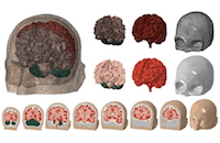 | This method is typically based on personal experience and contains the risk of leaving the patient with severe disabilities. During decompressive craniectomies, nerve fibers in the brain, known as axons, stretch, and run the risk of shearing, making this procedure a "last resort" for surgeons. Breakthroughs are being made in this area at the Stevens Institute of Technology, Stanford University, Oxford University, and the University of Exeter. Researchers have developed a workflow employing Simpleware software for medical image segmentation and generation of a Finite Element (FE) model of the brain to simulate craniectomies under different conditions. The use of these methods gives neurosurgeons insight into extreme tissue kinematics, enabling them to plan the shape and position of the craniectomy. More Information: synopsys.com/news |